Procedure for White Matter Parcellation:
Frontal Lobe White Matter:
Change "cerebral white matter" on every slice that has frontal lobe present on it to "frontal lobe white matter." This will, on some slices, for the time being, change some white matter that is actually temporal or parietal white matter to frontal lobe white matter. This will be corrected later on, however.
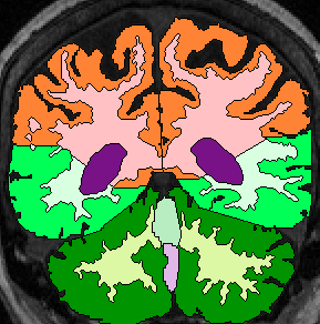
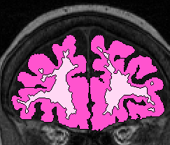 (fig 1)
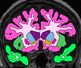
 Temporal Lobe White Matter:
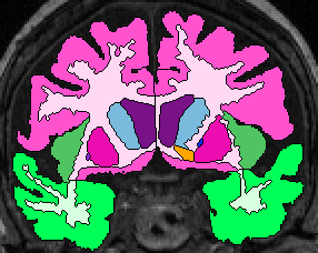
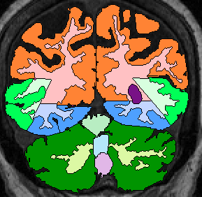
Anteriorly, simply relabel the cerebral white matter in the temporal lobe as temporal lobe white matter (fig 2). When the white matter becomes continuous from the temporal lobe to the frontal, draw a horizontal line laterally from the inferior Insula, then extract the temporal white matter, leaving it unlabeled for the time being (fig 3- this image show it labeled, however). Once the frontal lobe gray matter superior to the amygdala is gone, draw a straight horizontal line from one fronto-temporal junction (lateral sulcus) to the other and extract the temporal white matter (fig 4).
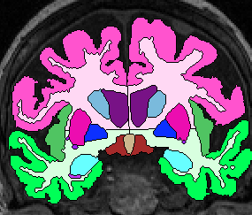
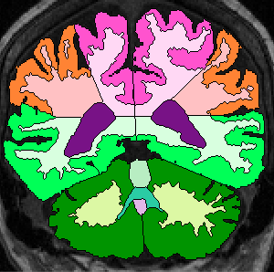
When parietal lobe shows up ventrally, draw a line from the medial parietal-temporal junction to the the lateral one(fig 5).
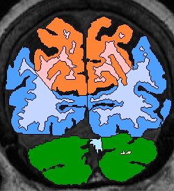
When occipital shows up inferiorly, extend the temporal boundry lines straight out until they intersect (fig 6).
Finally, label all unlabeled extractions as Temporal White Matter.
 (fig 2)
 (fig 3)
 (fig 4)
 (fig 5)
 (fig 6)
Occipital Lobe White Matter:
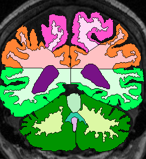
Anteriorly, extend parietal occipital gray matter boundary until it intersects the temporal boundary and extract this area (fig 6).
Posteriorly, extend boundry lines to intersect and extract occipital white matter (fig 7).
Finally, label all unlabeled extractions as Occipital Lobe White Matter
 (fig 7)
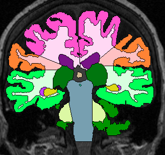
 Parietal Lobe White Matter:
Extend fronto-parietal gray matter boundary more or less straight to the CSF Third vents (fig 8). When third vent is gone, simply extend fronto-parietal boundary line to intersect with temporo-parietal boundary. Extract this parietal white matter area, but leave unlabeled for the time being.
When parietal gray matter shows up medially, extend the boundry lines to intersect. Continue to do this when occipital lobe appears (fig 7). At this point, most of the boundaries are already drawn.
Finally, label all unlabeled extraction as Parietal Lobe White Matter.
 (fig 8)
 (fig 9)
 (fig 10)