Segmentation: Nucleus Accumbens

General Description
In the area just above the orbital surface of the frontal lobe the head of the caudate appears to be continuous with the anterior part of the putamen. This region of continuity is the referred to as the nucleus accumbens.

The nucleus accumbens is bordered superiorly by the internal capsule, caudate, and putamen. It is bordered inferiorly by white matter, or in its most posterior extent by the subcallosal gyrus. Its medial border is the septal nuclei, or lateral ventricle. Laterally it is bordered by the putamen.

Because the nucleus accumbens is nearly impossible to see in a standard MRI, the CMA has come up with a convention for the segmentation of this structure.

Procedure

Segmentation
The outline for the nucleus accumbens is created using the intensity contour function. The outline is most often taken at the same time as putamen and/or caudate. The nucleus accumbens is isolated by separating the caudate from the putamen. As a rule, accumbens is not taken if anterior commissure is visible.

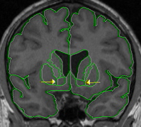
Part I - Caudate and putamen are discontinuous
The most anterior nucleus accumbens is taken in the first slice where both the caudate and putamen are present. When the caudate and putamen are not connected, an oblique line should be drawn from the inferior most tip of the lateral ventricle (where it meets the caudate) to the inferior most, medial tip of the putamen. This line will provide the superior border of the accumbens. The inferior border should be achieved using intensity contour, most often the same line used for the caudate.


Part II - Caudate and putamen are continuous
When caudate and putamen are connected, an oblique line should be drawn from the inferior most tip of the lateral ventricle to the inferior most tip of the internal capsule (the white matter area between the caudate and putamen). If the white matter tip is an "island" it should be connected with a line to the rest of the internal capsule and the division lines should be drawn with the "island" as the reference. Adjust the intensity contour to clearly see the full extent of white matter, to it's most inferior extent. This line will create the border between the caudate and the accumbens. From this point, a straight, vertical line should be drawn to provide the border between accumbens and putamen. The accumbens should end a slice or two in front of anterior commissure.

This area can be tricky in terms of order of extraction and method of achieving the conventions. It is helpful to play around to figure out your own style but we recommend the following procedure: extract caudate and putamen each with their own method and provide a temporary inferior border for each structure. Find borders for nucleus accumbens either with intensity contour or by manual drawing (often useful for the inferior border). Extract all three structures together from the outside, unextract the putamen and caudate, erase (using x) the temporary borders. This will give you a big "U" shape. Isolate the accumbens as described above, then unextract the "U" from the outside and re-extract each structure individually.

Labeling
The final outline should be labeled "accumbens area."

© 2005 Neuromorphometrics, Inc. All rights reserved.