The amygdala is located in the medial temporal lobe. It has a rounded shape and is situated anterior and superior to the hippocampus. Anteriorly, the amygdala borders the entorhinal/perirhinal temporopolar cortex. Superiorly, it borders the basal forebrain and the choroidal fissure. Medially, the amygdala borders the entorhinal cortex (in its anterior most tip) as well as the subarachnoid CSF of the medial temporal surface. Its lateral borders are the temporal horn of the lateral ventricle as well as (more rostrally) the white matter core of the temporal pole. Inferiorly, it borders with the entorhinal cortex (PHa), and more posteriorly, the hippocampus. Its caudal border is the hippocampus.

Procedure
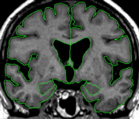
Sulci Lines
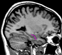
The amygdala is very difficult to see in the coronal view. For this reason, both the sagittal and axial views are very important in understanding the shape of the amygdala. The user should make an effort to learn the anatomy of the amygdala very carefully in these views. Furthermore, sulci lines will prove very useful in determining the borders of the amygdala in the coronal plane.
Sulci lines must be drawn in a different color than the one used to segment in the coronal view. Use the sagittal view to separate the hippocampus from the amygdala. Start with one side of the brain and move to approximately the most lateral extent of the amygdala. On this slice you will see the hippocampus, a large portion of the inferior lateral ventricle (ILV), and a small gray area that is the amygdala.


Drawing lines around the amygdala in the axial view (particularly inferiorly) and sagittal (also inferiorly) will help with the shape of the anterior amygdala in the coronal view.
Segmentation
The amygdala is segmented using a contour line and manual editing.
Part I - Anterior portion of the amygdala
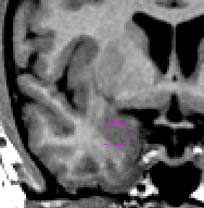
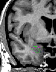
The amygdala begins when the cortex begins to look "puffy." If you've drawn amygdala sulci lines, these will start to appear on your first slice of amygdala. On this slice, check both the sagittal and axial views to make sure there is indeed amygdala on that slice. Using an atlas as a guide to where the first slice of amygdala appears, draw a circle in approximately this area. Then, using projection lines, check both the sagittal and axial views to edit your circle to include only the amygdala. It is most useful to check the middle of your circle to approximate the dorsal-ventral, medial-lateral extent of the amygdala. Then check your medial-lateral approximations in the axial view to make sure you really are only including amygdala in this outline. Note that on this first slice, the amygdala should not touch the cortex. These slices should be labeled "anterior amygdala."
The second slice of the amygdala should be segmented using the same methodology. The only difference is that the amygdala may touch the cortex on this slice. Also, it may be possible to see the lateral extent of the amygdala on this slice. If that is the case, use a contour line to accurately capture this border of the amygdala.
As you move posteriorly, the amygdala becomes easier to visualize. Use a contour line to give the general outline of the amygdala, then check the other views available to you to confirm this outline. When the amygdala is in its full extent, it is fairly easy to see in the coronal view. Remember that the amygdala has a very small wave that crests over the choroidal fissure. Refer to an atlas to see this wave more clearly. Alter the brightness of the screen so that you can adequately see the strip of white matter that separates the amygdala from the cortical areas.
When the ILV is first visible, it is likely that the hippocampus is present; check for your hippocampal sulci lines as well. To segment the amygdala in this area, use a contour line to define the hip-amyg area. Call up your sulci lines. Depending on the brain, you will see darker pixels that represent the ILV, or lighter pixels that represent the fimbria of the hippocampus (or both) along your sulci lines. If possible, draw a line separating the amygdala from the hippocampus along the dark pixels of ILV. Try increasing the contrast between black and white to better see this division. If after manipulating the brightness/contrast this line is not visible, draw a line that bisects your sulci lines. Then use projection lines to verify that your line really is the division between the hippocampus and the amygdala. When you are satisfied, unextract the hip-amyg outline, and extract the top portion as amygdala.
Part IV - Posterior portion of the amygdala
As you move posteriorly, the amygdala will become smaller as the hippocampal area increases. Continue to follow the procedure outlined above to separate the hippocampus and the amygdala. Continue to take the amygdala until it is no longer visible on the coronal view.
Labeling
Label the most anterior slice of amygdala as "amygdala anterior." Label all subsequent slices of amygdala "amygdala."