
General Description
The three parts of the brainstem include the midbrain (mesencephelon), the
pons, and the medulla. The most superior part of the brainstem is the midbrain
which continues behind the pons and down to the medulla. The more anterior,
superior part of the brainstem is the pons and the more posterior, inferior
part of the brainstem is the medulla, although there is some overlap. The
pons is an apple shaped structure, which sits on the anterior side of the
more stalk-like medulla. The medulla leads directly into the spinal cord.
The posterior border of the brainstem is the cerebellum, although cerebellum
and brainstem are present at the same time. The brainstem is bordered superiorly
by the diencephalon and inferiorly by the spinal cord. The superior colliculi
and inferior colliculi (seen as two bumps on top of the brainstem in more
posterior slices) are included as brainstem.
Segmentation Procedure
Part
I. Sulci Lines
Draw two sulci lines in the sagittal view to determine the superior/inferior
borders of the brainstem.
1. For the first sulci line, draw the superior brainstem line as a straight
diagonal line extending from the tip of the posterior commissure to the prepontine
fissure to mark the border between the pons and the midbrain. In coronal sections
this line will mark the superior border of the brainstem from the ventral diencephelon
(VDC).
 2.
The inferior brainstem line extends from the obex (bottom) of the fourth ventricle
across the width of the brainstem to the pyramidal decussation (bottom of the
pyramidal tracts). In coronal sections this line will mark the inferior border
of the brainstem from the spinal cord.
2.
The inferior brainstem line extends from the obex (bottom) of the fourth ventricle
across the width of the brainstem to the pyramidal decussation (bottom of the
pyramidal tracts). In coronal sections this line will mark the inferior border
of the brainstem from the spinal cord.
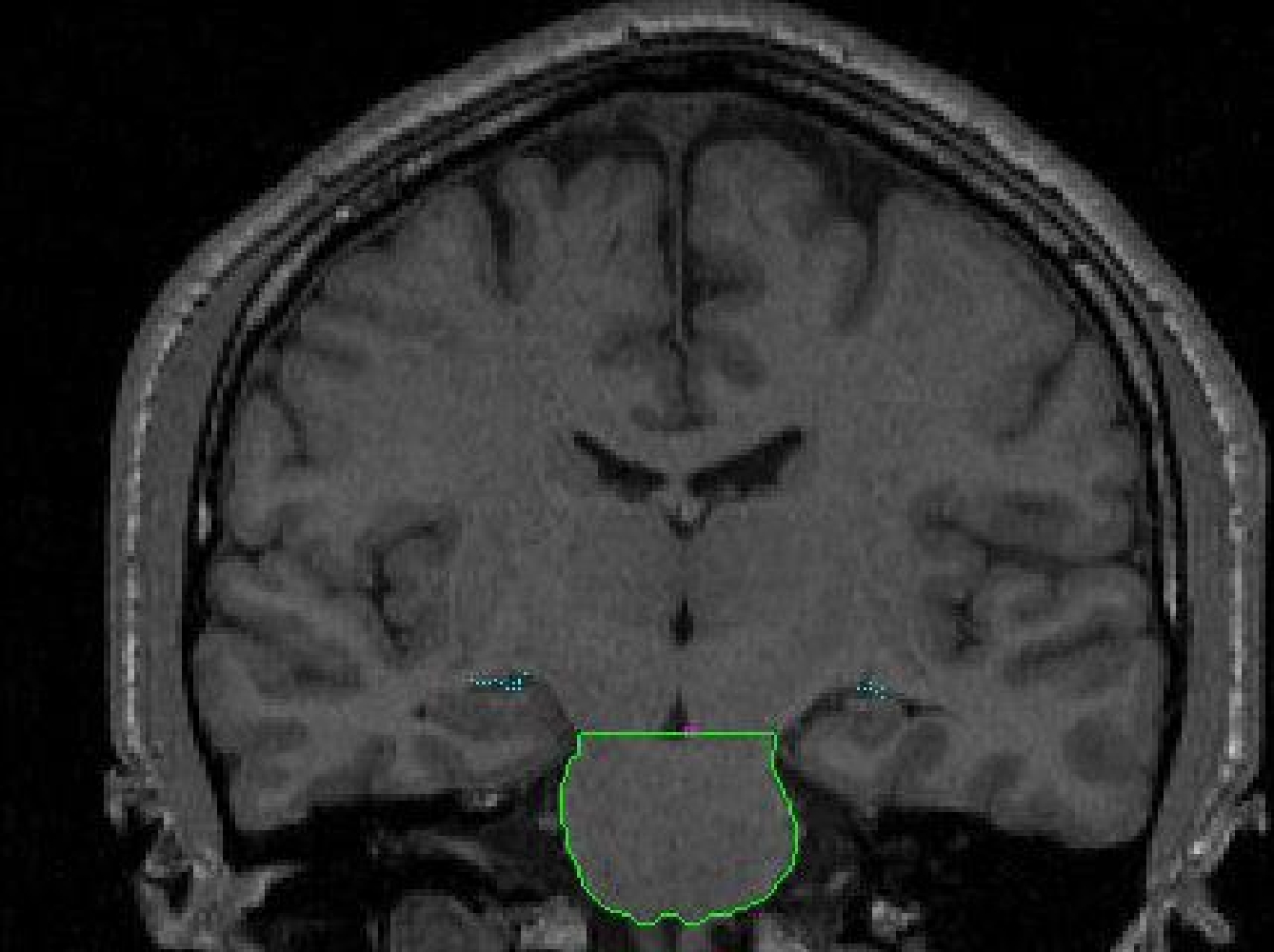
Part II - Anterior portion of the brainstem
1. Begin segmenting the brainstem when the first slice containing the brainstem is visible. Use the contour function (C) to create a contour that hugs the area of the brainstem. The draw function will also need to be used to connect parts of the contour that are not connected, or to exclude non-brainstem parts from the outline. It may be necessary to use the piece-wise contour method (V) on certain areas of the brainstem.
 2.
Before your sulci lines are visible, the interpeduncular fossa is used as a
reference for the superior border of the brainstem. The interpeduncular fossa
is an area of CSF between the cerebral peduncles, where the cerebral peduncles
connect with the brainstem. If the border between the peduncles and the brainstem
is not visible then use the interpeduncular fossa as the starting point and
draw a diagonal border from the fossa to the lateral extent of the brainstem
on each side.
2.
Before your sulci lines are visible, the interpeduncular fossa is used as a
reference for the superior border of the brainstem. The interpeduncular fossa
is an area of CSF between the cerebral peduncles, where the cerebral peduncles
connect with the brainstem. If the border between the peduncles and the brainstem
is not visible then use the interpeduncular fossa as the starting point and
draw a diagonal border from the fossa to the lateral extent of the brainstem
on each side.
The inferior border in the anterior extent (around the pons and medulla) is
visible and can be discerned with the contour function.
Many of the cranial nerves appear in the area of the brainstem. Cranial nerves should be excluded from the brainstem outline. Many arteries or veins also appear around the brainstem, these should be excluded from the outline. For example, the basilar artery appears in the anterior slices of the brainstem and when discernible it is excluded from the outline of the brainstem.
 Part
III – Superior brainstem lines appear
Part
III – Superior brainstem lines appear
As soon as the superior brainstem line appears, it will be used as the marker
for the division between the VDC and the brainstem. The two structures are
separated by drawing a line which bisects the vertical sulci line (appearing
as a dot), such that everything above it will be labeled VDC and everything
below it brainstem.
Before the appearance of the cerebellum, there is a dramatic change in the
lateral extent of the brainstem. The more anterior of the slices in this region
will contain a uniformly colored structure which is extracted and labeled as
part of the brainstem (there is no cerebellum present at this point). More
posteriorly, one slice before the cerebellum first appears, the lateral extents
of the brainstem are segmented separately to exclude the middle cerebellar
penducles.

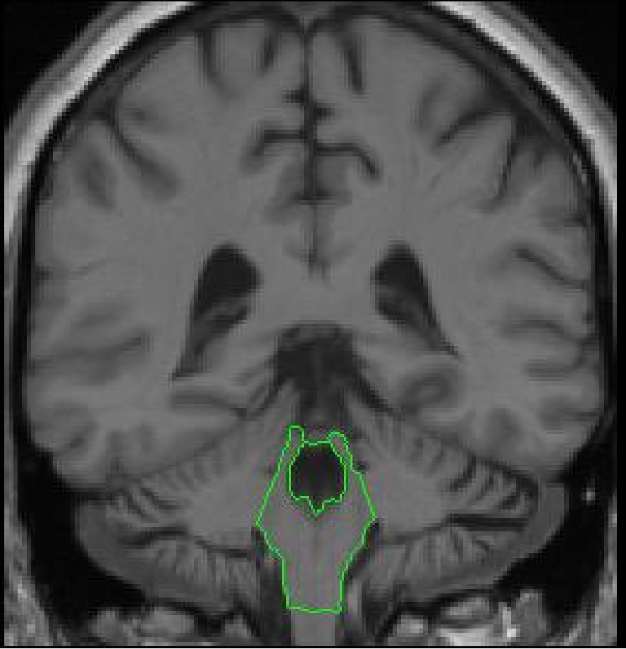
Part IV - Cerebellum appears
At its anterior extent, the cerebellum shares its medial borders with the brainstem.
The lateral extremities of the brainstem are no longer taken as part of the
brainstem outline. They are extracted separately as cerebellar exterior and
as cerebellar white matter. With the contrast increased it is easy to see the
division between cerebellar white matter and brainstem. This division can be
manually drawn in or it may be possible to use the contour function. The next
slices will have cerebellum present and these lateral extremities will again
be extracted as cerebellar white matter.
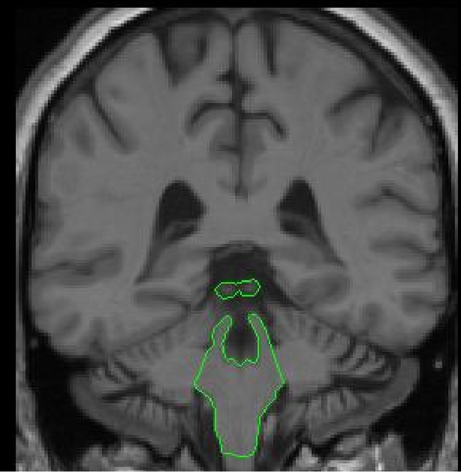
Part V - The colliculi appear
In the more posterior slices when the superior brainstem line is no longer
visible, it is necessary to draw in the superior and inferior colliculi manually
or with the use of the contour function. There may be a slice or two where
the colliculi are not yet attached to the brainstem, in this instance extract
them as a separate outline which will still be labeled brainstem.
In this area be careful to exclude the pineal gland from the volume of the
brainstem.
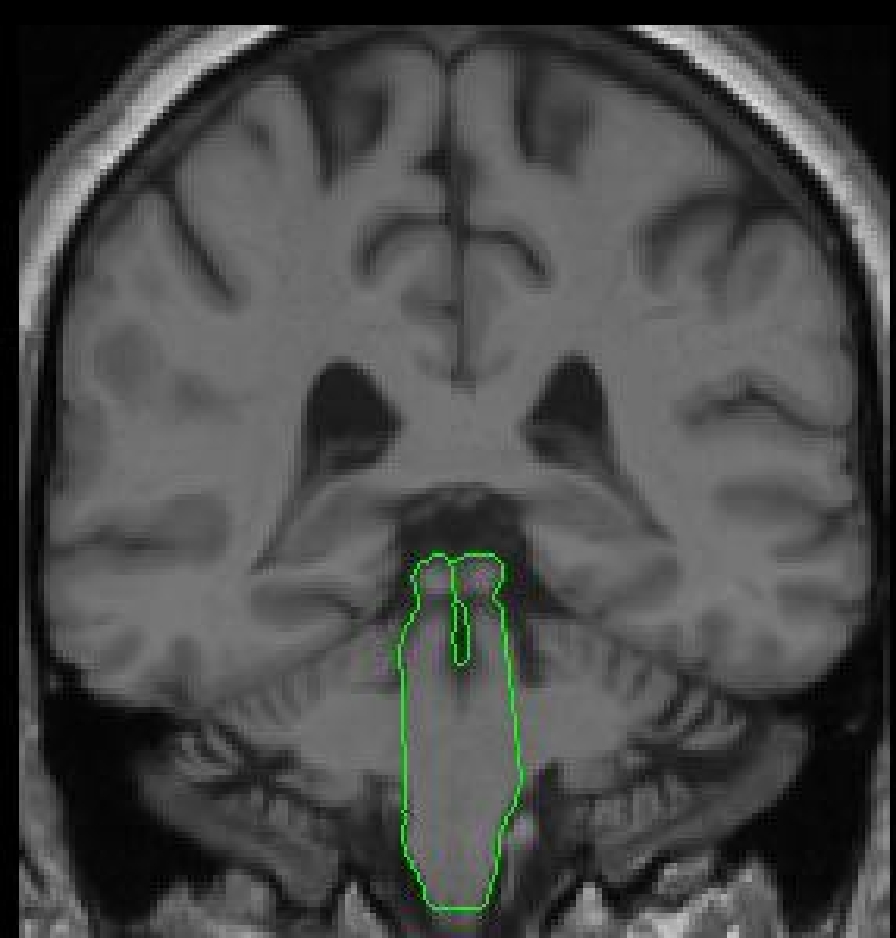
The inferior border in the more posterior extent (where the medulla is connected
to
the spinal cord) depends on the inferior brainstem line drawn for the inferior
border of the brainstem. Draw a line that bisects the inferior brainstem line
(appearing as a dot). Everything above this line will be brainstem, and everything
below it will be spinal cord and considered outside of the brain.
Be sure to always attach the 4th ventricle' to the brainstem exterior to exclude it from the volume of the brainstem.
Labeling
 The
final outline should be labeled "brainstem."
The
final outline should be labeled "brainstem."
© 2005 Neuromorphometrics, Inc. All rights reserved.