The cerebral white matter is comprised of any area of the brain with a high concentration of axons covered in myelin. Because myelin is made of lipid, it has a white appearance in MRI scans. During segmentation the white matter is separated from the cortex and subcortical structures using the histogram function. There is a different white matter parcellation program used to divide the white matter into its constituents.
Procedure
Segmentation
Cerebral white matter is extracted with the histogram method, as well as some manual drawing.
Create a histogram using the entire white matter-containing area as your "histogram box." Select the intensity which falls between the peak which represents cortical gray matter and that which represents white matter. This will generate your white matter contour line. Use a separate histogram for each side of the brain, and for the temporal lobes. When the white matter is continuous between the temporal and frontal lobes, you should be able to use one histogram for the entire hemisphere.
If the white matter appears continuously on the MRI scan, but your histogram does not give you a continuous outline, manually draw in a strip a white matter between the two lobes. Also, if the two lobes are connected (share the same exterior outline) the white matter must be continuous.
By convention and by anatomy, cerebral white matter should never extend through the cortical ribbon. Radiologically speaking, an image may be produced in which partial voluming contributes to the illusion that the white matter extends to the edge of the cortex, though anatomically, this does not occur in the normal brain. Manually edit as necessary to ensure the white matter does not touch the cerebral exterior along the edges of the brain. White matter does extend to the medial exterior border where the corpus callosum is located. This should be apparent with your histogram, if this is not apparent, manually edit to include all of the corpus callosum.
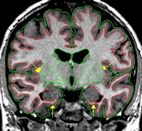
Be sure to have continuous white matter lateral to the putamen and below the hippocampus. If necessary, manually draw these borders.
Also, if a temporal lobe connects to a frontal lobe, then white matter must exist along that connection. If necessary, manually draw in a thin strip of white matter.
To be sure that you have extracted only white matter, click on the line believed to be white matter and it will turn white.
In the most anterior and posterior extents of the brain, the histogram you generate for the white matter will look different than it does for the rest of the brain. You will probably see three peaks: one small peak on the right, one large peak in the middle, and one smaller peak on the left. In this instance, create a contour by dragging your mouse from the larger middle peak to the smaller left peak (it is often helpful to expand the histogram with the third mouse button). It may also be the case that the best fit contour is between the middle peak and the last visible peak on your histogram (no matter what its height). Remember that it is unlikely you will have white matter on the very first and last slices of the brain.
Depending on the scan, you may witness a large extent of "drift." This term means that the intensity of the white matter is variable from one area to the other. For example, the white matter at the top of the image is much brighter than the white matter at the bottom of the image. In such cases, it may be necessary to piece together two white matter histograms. Draw a line between the top and bottom halves of the hemisphere. Extract the top portion and create a histogram. Clip the ends of the resulting contour at the point where it enters the bottom half of the hemisphere. Turn the contour yellow with the "v" function and remove stray lines. Then, using the histogram method, create a contour for the bottom part of the brain. Extract the two contours from the outside, and then the inside. Make sure you've completed all necessary manual editing.
On some slices, the fornix is not part of your cerebral white matter outline because the lateral ventricles create a division between the corpus callosum and the fornix. Extract this small area of fornix and label it as white matter.
Labeling
Label this outline as "cerebral white matter."