Cerebral Exterior
General Description
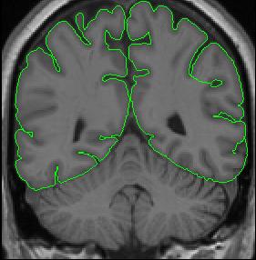
The cerebral exterior is the border between the subarachnoid CSF and neural tissue (e.g. the first layer of cortical neurons), and should correspond to the pia mater. Thus, the cerebral exterior separates brain from non-brain, cerebrum from cerebellum, and divides the brain in to its two hemispheres. Your outlines should not include anything that is not brain (e.g. dura mater, other meninges, etc.). For our purposes, optic chiasm is considered to be outside of the brain, and therefore excluded from exterior outlines. To determine what is and what isn't brain, it is useful to check the other two views (sagittal and axial). Once you've determined what is and isn't brain, use the draw tool to make the appropriate corrections on your exterior outline.
Segmentation Procedure
The exterior is defined using the intensity contour method and manual drawing.
- Increase the brightness of the image (use middle mouse button) to see the actual extent of the cerebral hemispheres. If the white matter begins to bleed into the gray matter, you have gone too far.
- Create a contour using the intensity contour function (press c) that is somewhat larger than the exteriors. Then, adjust the contour until it fits tightly around the hemispheres, making sure you don't exclude any gray matter from your outline. If when you generated your outline using the contour function you have a small contour inside the brain that represents a sulcus (e.g. the Sylvian fissure), you must connect this small contour with your exterior by tracing along the sulcus in the image.
- Be sure to exclude things that are not brain such as meninges and blood vessels.
- Return the brightness to normal intensity by decreasing the brightness (with middle mouse button). Complete your exterior outlines by manually drawing lines to complete the gaps remaining in your contour outline.
- The exterior line will need to be tighter in some areas. Specifically, make exteriors tighter around the hippocampus and amygdala, so as not to include vessels in that area.
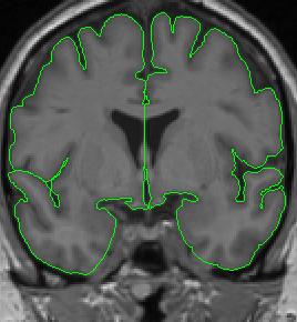
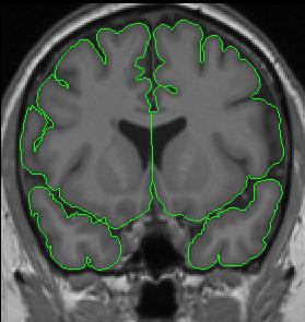
The cerebral hemispheres are extracted independently, and their division, is most clear in slices where they are completely separate.
When corpus callosum is present, it is necessary to separate the hemispheres by manually drawing along the midline.
Anteriorly, when the temporal lobes are present but not connected to the frontal lobes, the temporal lobes are extracted separately from the frontal lobes. Thus, you will have four separate outlines that make up the cerebral exteriors.
At the fronto-temporal junction, if the contour encompasses the entire hemisphere but the white matter between the lobes is not continuous, it is necessary to separate the frontal and temporal areas. Each hemisphere and lobe should then be extracted independently.