Segmentation: Fourth Ventricle
General Description
The fourth ventricle is located between the brainstem and the cerebellum. Its
anterior border is the brainstem. Laterally and posteriorly it is bordered
by the cerebellum. Its posterior border (above the cerebellum) is the midbrain
tectum (superior and inferior colliculi). The cerebral aqueduct in included
as part of the fourth ventricle.
Segmentation Procedure
The histogram function is usually recommended for extraction of the fourth
ventricle. Depending on which region of the fourth ventricle you are looking
at, the box drawn for your histogram will contain CSF from the ventricle
and either cerebellar white matter, cerebellum gray matter, brain stem, or
some combination thereof. The intensity contour method and manual drawing
are also employed.
 Part
I - cerebral aqueduct
Part
I - cerebral aqueduct
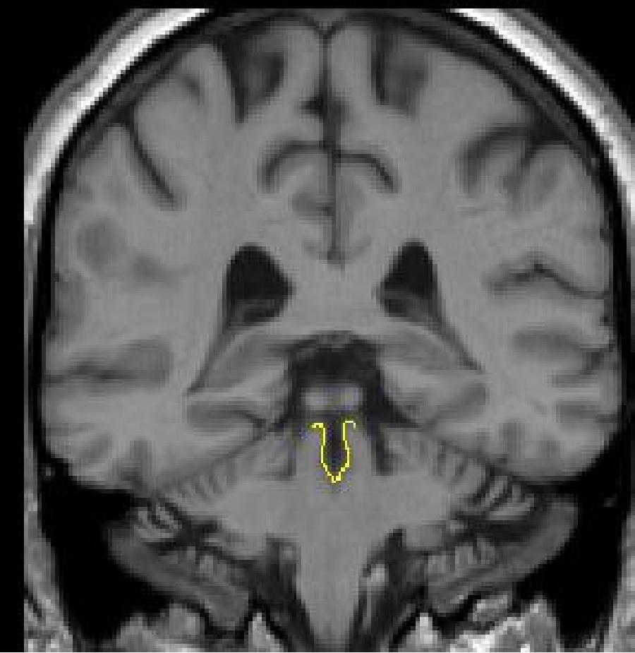
The aqueduct first appears just under the posterior commissure. A histogram
should be taken between the CSF of the aqueduct and the brainstem. However,
because there is so much partial voluming in this area, the histogram will
likely be modified using an intensity contour line. The dorsal border of the
fourth ventricle will have to be drawn manually. Continue to use a histogram
for the remainder of the aqueduct, modifying as necessary with the intensity
contour function.

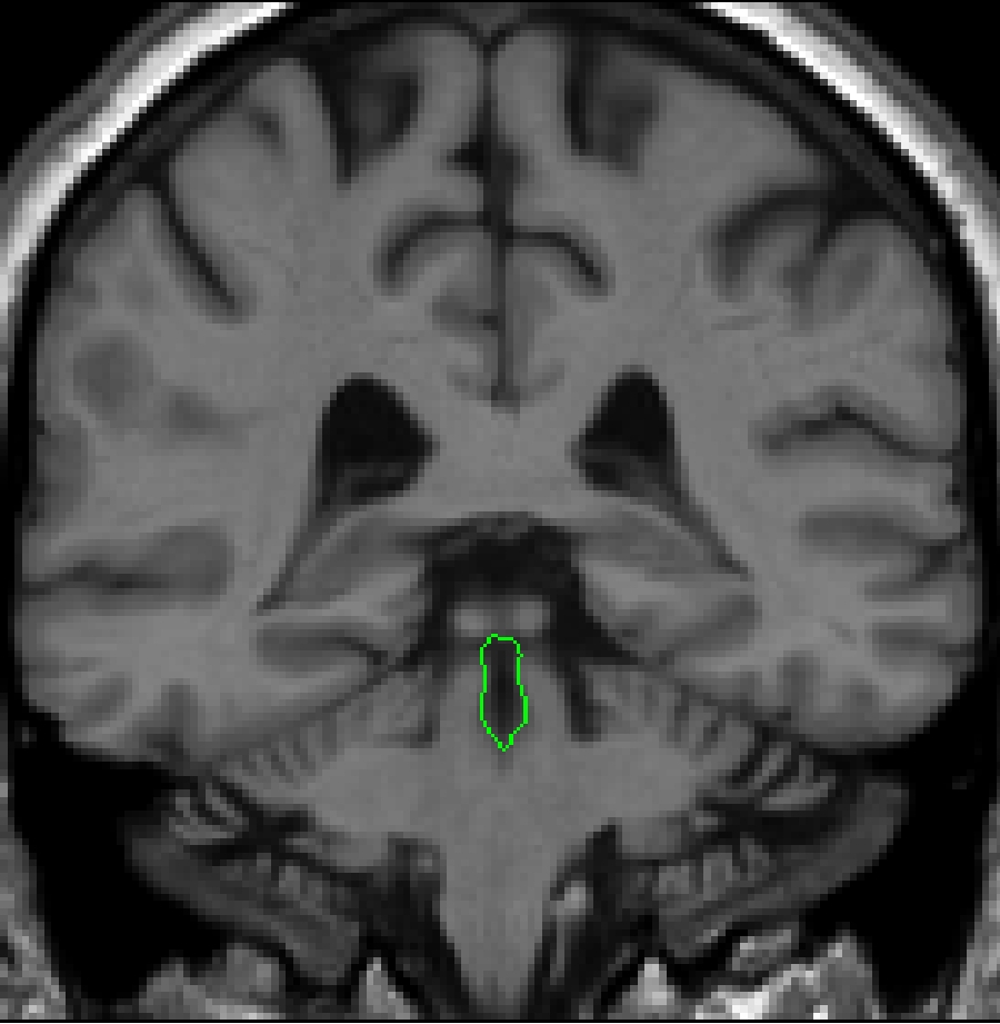
Part II - fourth ventricle in the brainstem
As you move posteriorly, you will begin to see the actual beginning of the
fourth ventricle. The small circle that is the aqueduct will begin to elongate.
Continue to use the histogram method; draw your box between the CSF of the
fourth ventricle and the surrounding brainstem tissue. Modify as necessary
with the intensity contour function. As the fourth ventricle continues posteriorly,
it will start to widen. A histogram should be taken between the CSF of the
fourth ventricle and the surrounding brainstem tissue. Often this histogram
will not yield the dorsal border of the 4th
 ventricle.
Brightening the screen will enable you to see this border. It should be drawn
in using the draw function, and then attached to the contour given by your
histogram.
ventricle.
Brightening the screen will enable you to see this border. It should be drawn
in using the draw function, and then attached to the contour given by your
histogram.
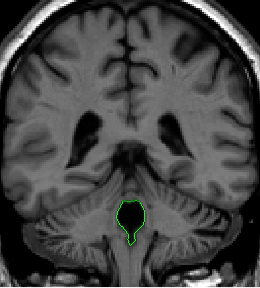
Part III – fourth ventricle in the brainstem and cerebellum
 As
the 4th ventricle is surrounded by cerebellum white matter, multiple histograms
will yield the most accurate fit. Generate a histogram from a box containing
CSF of the fourth ventricle and the cerebellum white matter. The only part
of the contour that you want is that between the cerebellar white matter
and the CSF of the fourth ventricle. Now generate the rest of the outline
with the histogram method.
As
the 4th ventricle is surrounded by cerebellum white matter, multiple histograms
will yield the most accurate fit. Generate a histogram from a box containing
CSF of the fourth ventricle and the cerebellum white matter. The only part
of the contour that you want is that between the cerebellar white matter
and the CSF of the fourth ventricle. Now generate the rest of the outline
with the histogram method.
The box for your second histogram should contain equal amounts of CSF from
the fourth ventricle and the brainstem. The generated contour will accurately
define the border between the fourth ventricle and the brainstem.
Part IV - fourth ventricle in the cerebellum
 When
the fourth ventricle is no longer surrounded by brainstem, it appears between
cerebellum gray and white matter. Two histograms should be used for this
outline: one between the CSF and the cerebellum white matter, and the second
between the CSF and cerebellum gray matter.
When
the fourth ventricle is no longer surrounded by brainstem, it appears between
cerebellum gray and white matter. Two histograms should be used for this
outline: one between the CSF and the cerebellum white matter, and the second
between the CSF and cerebellum gray matter.
In its most posterior extent, the fourth ventricle will appear as two separate
circles in each cerebellar hemisphere. The most accurate means to extract these
structures is to do two separate histograms for each cerebellar hemispheres
(CSF - white matter; CSF - gray matter). As with the most anterior extend of
the 4th ventricle, modifying this
estimate with the contour line may be necessary.

Labeling
Both the cerebral aqueduct and fourth ventricle are labeled as "fourth
ventricle.
©
2005 Neuromorphometrics, Inc. All rights reserved.