| General Description What we refer to as the inferior lateral ventricle (ILV) is actually the temporal horn of the lateral ventricle. This structure is located in the temporal lobe, and throughout its course it will change significantly in shape. The inferior lateral ventricle is extracted so as to exclude it from the hippocampus and amygdala outline; it is not considered a reliably extracted structure that is used for its own morphometric analysis. Most researchers combine the volume of the inferior lateral ventricle with what was previously described as the lateral ventricles when doing morphometric analysis on the lateral ventricles. Because of this, there are many methods that can be used to extract the ILV. | 
| Procedure Segmentation  
 | 
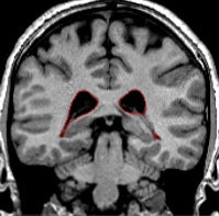
|  | Anteriorly, the ventricle will appear more as a curved structure which follows the rounded lateral edge of the hippo-amyg area. Moving posteriorly the inferior lateral ventricle will comprise the border between the hippocampus and amygdala. Create an intensity contour line for the ILV. Your contour line will approximate the value of the lateral ventricles and/or the part of the tightened exterior that borders the hipp/amyg area. Once complete, extract this outline from the outside, and then the inside. 
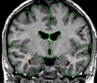
It is also acceptable and more accurate to use a multiple-peaked histogram for the ILV, when accuracy is important. It is possible to draw one box that contains all three of the structures that make up the ILV borders (CSF, white matter, hipp/amyg), and then use the corresponding peaks to create the ILV outline. The first to second peaks will represent the CSF to hipp/amyg (gray) averaged intensity, and the second to third peaks represent the hipp/amyg (gray) to white matter averaged intensity. Use the "v" function to create the outline. | 
| Part II - Medial portion of the ILV Moving posteriorly, when the amygdala is gone or almost gone, it may be difficult to determine the medial extent of the inferior lateral ventricle. Depending on the brain, it may appear as the inferior lateral ventricle is continuous with the exterior outline. There is in actuality a small membrane called the tele choroides of the lateral ventricle that separates the inferior lateral ventricle from the outside of the brain. Brightening the screen may help to see this thin membrane. If it is not possible to see this border, discretion must be used. The inferior lateral ventricle extends medially to the subiculum, and not past. Once you have made a decision as to the most medial extend of the inferior lateral ventricle, remain consistent with this decision through the posterior course of the brain. As you continue to move more posterior, this will no longer be an issue. | 
| Part III - Posterior portion of the ILV Toward its posterior endpoint, the ventricle will appear as a small circle which is adjacent to the ventral-lateral corner of the hippocampus. The course of the inferior lateral ventricle is sometimes interrupted, and may be absent for a slice or two but then reappear. Use of the projection lines will verify the extent of the ventricle at its anterior and posterior limits. At its posterior endpoint, the inferior lateral ventricle will become continuos with the lateral ventricle outline. | 
|  | 
Labeling 
This outline is labeled as "inferior lateral ventricle."
© 2005 Neuromorphometrics, Inc. All rights reserved.