
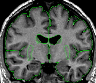
The putamen is a subdivision of the lenticular nucleus (the other division is the pallidum). The lenticular nucleus from the axial view resembles a rounded triangle that is divided into two major sections. The putamen is the lateral part of this triangle.The putamen starts small and ends small in the coronal view. The putamen quickly grows to its greatest size in the middle and in the medial posterior portion it closely resembles a goldfish shape. The putamen lies lateral and partially anterior to the thalamus. It is bordered laterally, superiorly, and inferiorly by white matter.
The putamen is usually bordered medially by the pallidum. When the pallidum is not yet present or has already disappeared the putamen is bordered medially by the internal capsule.
Procedure
Segmentation
The putamen outline is created using the intensity contour function and manual drawing.
Part I - Putamen
Begin by generating a partial outline that you think fits a portion of the putamen, then using the erase function, clip this line where it no longer fits the putamen. Use the "v" function to "save" the line. Create another line with the intensity contour function to generate the rest of the outline. Clip this line where it joins your first line. Use the "v" function again to save this new line. Now extract the completed putamen from the outside, and then extract from the inside.
It is important not to include the claustrum in the putamen; this is the strip of tissue bordering the lateral edge of the putamen.
Often the putamen can be extracted at the same time as the caudate especially in the area where they are connected by the nucleus accumbens. The histogram of the caudate, in many cases, is close to what you want for the putamen as well. After extracting the caudate, before deleting the remaining red lines, you can often go immediately into the intensity contour function and adjust them to fit the putamen. This should be done separately for each hemisphere.
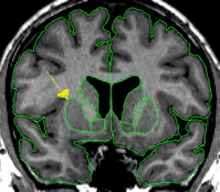
It is important not to include all of the "fish tail" of the putamen, this is the portion that appears to extend to the amygdala. This is not included as part of putamen; there should be a strip of white matter between amygdala and putamen.
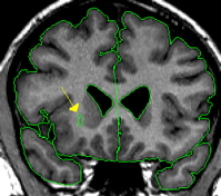
There is often a blood vessel near the inferior border of the putamen, this should not be included as part of putamen. The vessel should be extracted separately using a contour line and labeled "vessel." The vessel will serve as at least a portion of the inferior border.
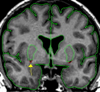
The final outline should be labeled as "putamen," and any blood vessels extracted should be labeled as "vessel."