General Description
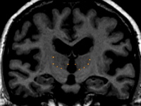
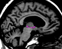
The thalamus is a large egg-shaped nuclear mass present in both hemispheres at the midline. The two thalami are separated medially by the third ventricle, cerebro-spinal fluid (CSF), or the cerebral exterior midline. They are bound laterally by the internal capsule. Each thalamus extends anteriorly to the interventricular foramen (foramen of monroe), and posteriorly the thalami overlap the midbrain and are bordered by CSF. The inferior border is the hypothalamic fissure, or the hippocampus in the most posterior extent. Superiorly the thalamus extends to the transverse cerebral fissure (TCF), lateral ventricle, white matter, or in the anterior portion, the caudate.
Procedure

By drawing many sulci lines in the dorsal axial view, the mid axial view and the ventral axial view of the thalamus you can obtain a skeleton of the outline of the thalamus produced by "dots" marking the sagittally drawn sulci in the coronal view. This gives you a guide to find the best fit contour line.
Part I - Anterior portion of the thalamus
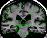
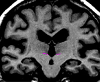
The anterior most extent of the thalamus appears after a few slices of the anterior VDC and is usually just posterior to the anterior commissure. In this region the thalamus appears as a thin sliver that gradually grows bigger and rounder as you move more posterior. It is bordered medially by the third ventricle and the dorsal border extends superiorly past the dorsal border of the third ventricle but not quite to the lateral ventricle or caudate. As you move more posterior the thalamus begins to touch the lateral ventricle, white matter, and in some cases a little of the caudate.
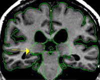
Part II - Posterior portion of the thalamus
The posterior extent of the thalamus is referred to as the pulvinar. The pulvinar extends posteriorly past the VDC and is located just superior and medial to the hippocampus. In this region the pulvinar is egg shaped and grows smaller as you move to the most posterior extent.
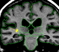
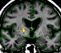
The dorsal medial border must be drawn in in order to exclude the fornix and separate the thalamus from the tissue between the ventricles. This is achieved by manually drawing in the border along the foramen of monroe or, if the foramen of monroe is not visible, from the bottom corner of the lateral ventricle to the medial cerebral exterior line or third ventricle.
The inferior border of the thalamus can be drawn by connecting the "dots" that result from the sagittally drawn sulci lines in the coronal view.