Probabilistic Brain Atlas
Embed this atlas into your software to localize or identify neuroanatomy
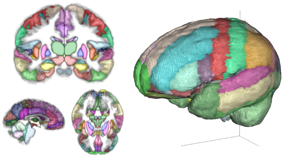
We do millimeter-resolution manual labeling of MRI brain scans and have used our database of individually-labeled scans to create the atlas shown above. Colors indicate the neuroanatomical region and grayscale gives the probability. The figure shows only maximum probability regions but the atlas gives all probabilities.
This atlas can both identify and localize anatomy for any kind of brain imaging, sensing and stimulation. The subject’s age, sex, handedness, etc. can be “dialed in” to make a subject-specific atlas and it can be registered to the subject using internal and external landmarks. If you already have an MRI or CT scan of your subject, this will help find and identify regions of interest: click on a point and the atlas provides a sorted list of probable regions. Without an MRI or other scan, this atlas will provide the most likely location of a neuroanatomical area. Unlike a template of a single subject, our results provide an indication of the variation of the living human brain.
Neuromorphometrics is the only company working on creating such a “gold standard” database. Our plan is to constantly re-analyze our labeled scans to improve it by fixing problems, adding regions of interest, in addition to labeling more scans to increase the age range and diversity of subjects. Our current database of labeled human MRI brain scans is already the largest, most comprehensive, and most consistently measured collection that is available anywhere (114 scans with subject ages from 5 to 96 years old). Yet it is only a start: we’re aiming to capture the variation of all brains on the planet. We label the entire brain and divide the cortex into regions based on gyral and sulcal landmarks using 1) the “General Segmentation” Protocol defined by the MGH Center for Morphometric Analysis (see here), and 2) the brainCOLOR Cortical Parcellation Protocol (from here).
Neuromorphometrics spun out of the Harvard/MGH Center for Morphometric Analysis with the help of SBIR grants from the NIH and we have been providing brain-labeling services for almost two decades. It used to take two weeks to label a single brain. Now, we do it in 2-3 days and we do a far better job with a lot more regions of interest. As we developed software and anatomical protocols with an eye toward reliability and reproducibility, we decreased the time required for labeling by concentrating on work-flow optimization and adding automation. One can’t do automated labeling without knowing how to do it manually first. Now we’re working on probabilistic atlases for neuroanatomical localization and identification. In the future, we will include white matter anatomy by labeling diffusion-weighted scans.
We will license our data to you directly or you can provide our data to your customers as a part of or as an add-on to your device or system. In addition to the atlas, we can also provide the individually labeled scans and we’ll also be happy to label scans that you provide.
For more details, see Neuromorphometrics.com, call +1 617-776-7844 or email Andy@Neuromorphometrics.com.