Lateral Ventricles
General Description
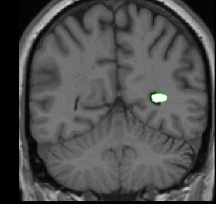
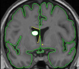
The lateral ventricles are bilateral C-shaped structures that extend through all four lobes of the brain. They are filled with cerebral spinal fluid (CSF), and for this reason appear black on the MRI scan. There are five different parts to each lateral ventricle: the anterior horn (in the frontal lobe), the body (in the frontal and parietal lobes), the posterior horn (extending in the occipital lobe), the inferior horn (in the temporal lobes), and the atrium (where the body, inferior horn, and posterior horn meet). For the purposes of segmentation, we consider all parts except for the inferior horn as lateral ventricle. The inferior horn is labeled as "inferior lateral ventricle" and its method of extraction is described elsewhere.
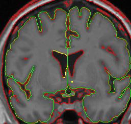
The lateral ventricles are bordered anteriorly by white matter. As you move posteriorly, the lateral wall of the ventricle is bordered by the caudate, and medially by white matter. Moving posteriorly, the lateral ventricles may appear as if they are connected along the midline. They are actually separated by the septum pellucidum. At this point the bottom wall of the lateral ventricle is bordered by thalamus. As you continue to move posteriorly towards the atrium, the thalamus no longer borders the ventricle; hippocampus becomes the medial border. Caudate still comprises the medial border of the ventricle, but it is difficult to visualize on the MRI scan. As you move past the atrium, the lateral ventricle is surrounded by white matter.
Segmentation Procedure
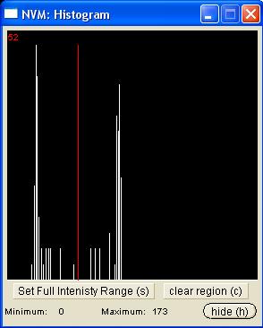
The histogram method is used to create outlines for the lateral ventricles. One histogram is needed to determine the CSF/white matter border, and another is used to define the CSF/gray matter border. A separate circle and histogram should be generated for each ventricle.
Part I - Anterior portion of lateral ventricles
- Draw a small circle that is half-way in the CSF from the center of the ventricle and and half-way in the white matter from the corpus callosum.
- Extract the box ( press "e")
- Generate a histogram (press "f")
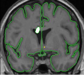
- Extract the lateral ventricle outline from the outside (press "e")
- Clean the stray pixels and extract from the inside of the ventricle by pressing "I"
Part II- Lateral ventricles with caudate present
When the caudate is present two histograms are needed to define the two different borders of the ventricle.
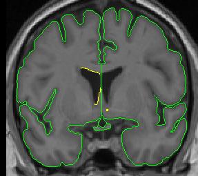
- Draw the first circle and create a contour for the CSF/white matter border (see above). This contour line is not a good measure of the border between the caudate and the CSF, so a separate line will need to be created for that section.
- Clip the two ends of the CSF/white matter line where the caudate lies. For the purposes of the lateral ventricles, we consider the thalamus white matter, meaning its border with the lateral ventricle is the same contour line as the CSF/white matter line.
- Use the "v" function to "save" the line.
- Create a line for the CSF/caudate border. Your histogram circle should be half-way in the caudate and half-way in the CSF.
- You may need to manually connect your "saved" contour to the new contour. By convention, in cases in which the caudate is present, include the most inferior extent of the CSF/white matter border as the lateral ventricle border, even if that necessitates drawing a short line from it to the CSF/gray matter border.
Part III - Posterior portion of lateral ventricle
Make sure the lateral ventricle is actually a ventricle, rather than a deep sulcus. To do this, look in other planes using the projection lines. There should be white matter between ventricle and the gray matter. Lateral ventricle can disappear for a few slices, and then re-appear.